The Oral and maxillofacial surgerydeals with the treatment of diseases of the mouth, face and neck, and related craniofacial structures.
There is great pathological diversity at this regional level, which is why oral and maxillofacial surgery combines infectious-inflammatory, traumatic, tumoral and congenital or acquired malformation.
Surgical techniques in oral and maxillofacial surgery
Reconstructive surgery: It is dedicated to the anatomical restoration of the bone structure and the reconstruction of soft tissues. The objective is that the intervened zone recovers its normal structure. A very common example is dental rehabilitation with implants.
Plastic surgery: The most common treatment is to correct facial malformations. It also covers other procedures such as skin-related treatments or facial implants.
Regenerative surgery: It combines the procedures of regeneration of tissues and treatments with stem cells. The periodontal area is one of the most treated.
Types of oral and maxillofacial surgery interventions
The field of oral and maxillofacial Surgery comprises:
Surgery and oral medicine
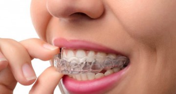
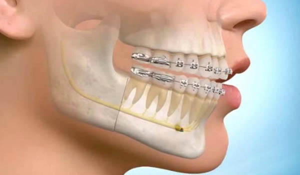
Interventions that are performed within the oral cavity. It includes the surgical extraction of the molars and other teeth, also the periapical surgery, excision of cysts and benign tumours of the maxillary, periprosthetic surgery, placement of dental implants, plasty of braces and mucogingival surgery. This type of surgery is done in ambulatory regimen and under local anaesthesia.
Facial injuries
The blows or traumatisms that happen on the face usually have some special characteristics different from those that occur in other areas of the body. The reason is that they can cause alterations, both from a functional and aesthetic point of view, as well as problems of vision, respiration or chewing.
Tumours in the mouth, face or neck.
The surgery of oral tumours represents one of the most important areas of the specialty, due to the complexity of the interventions and their repercussion in the life of the patient.
Together with other surgical specialists, the maxillofacial surgeon is an important medical figure in the approach to tumour pathology.
Facial alterations
This oral surgery is also known as orthognathic surgery. This is the set of surgical procedures that have the purpose of correcting alterations in the shape, position or size of the elements of the patient’s face to obtain facial harmony. Dentofacial deformities may be congenital, arise in people’s growth phase, or result from trauma.
Pathology of the salivary glands
Maxillofacial surgery is frequently used to treat those pathologies of the inflammatory or salivary glands.
Pathology of the temporomandibular joint
Temporomandibular dysfunction syndrome causes pain and limitations in mandibular dynamics. In addition, the temporomandibular joint (TMJ) can be affected by some degenerative diseases or trauma.
There are multiple treatments to treat problems in the TMJ and its associated musculature, among which surgery is included.
Facial aesthetic surgery
Maxillofacial surgery intervenes in the field of aesthetic surgery of the face, along with other specialties: plastic surgery, dermatology, ophthalmology and otorhinolaryngology.
Diagnostic tests in oral and maxillofacial surgery
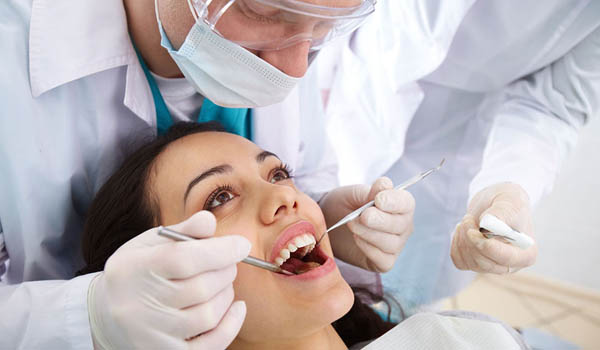
In the planning of the surgical intervention it is necessary to carry out a series of diagnostic tests in order to obtain a complete vision of the needs and characteristics of the patient.
General Radiology. X-rays are essential tests for diagnosing different types of facial bone lesions, such as malformations or malocclusions.
Biopsies . These tests may be necessary to determine if the lesions of the oral mucosa are due to a tumoral process.
Orthopantomography. It is a panoramic X-ray of the two jaws in order to have a complete image of them in a single plate. With this impression you can analyse the position of the teeth with more precision to plan the surgical procedure.
Magnetic resonance imaging . Very useful in the treatment of the temporomandibular joint, since it allows to fully visualize the articular disc.
3D studio. It is used to obtain a three-dimensional image of the area on which it is necessary to intervene surgically for a better planning of the process.